
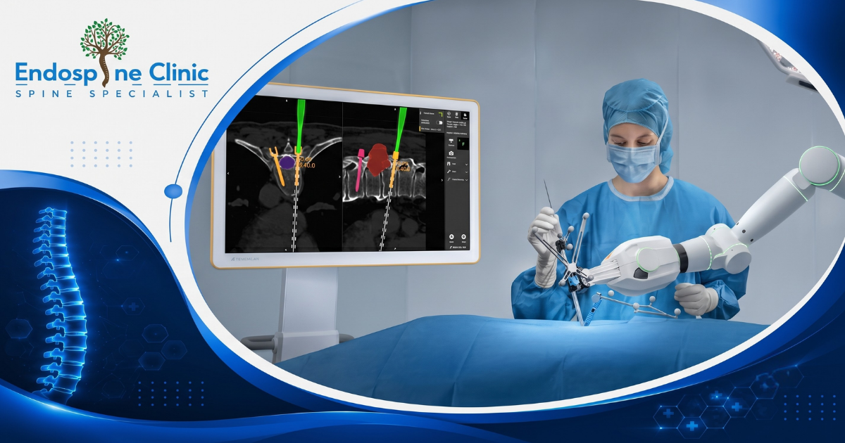
From X-rays to 3D maps: how technology guides spine surgery is revolutionizing the way back pain and spinal disorders are treated today. If you are struggling with chronic back pain, sciatica, or complex spinal conditions, the idea of undergoing surgery can be daunting. However, the medical landscape has completely transformed over the last decade. Gone are the days when surgeons relied solely on basic 2D imaging and large open incisions.
Today, advanced surgical navigation systems, robotic arms, and real-time 3D mapping are making spinal procedures safer, more accurate, and far less invasive. For patients seeking relief, understanding these advancements is the first step toward a pain-free life. Whether you are exploring options across India or looking for the best spine surgeon in Vadodara, knowing how these tools work will help you make an informed decision about your health.
In this comprehensive guide, we will explore the massive leap from traditional X-rays to modern 3D navigation. We will break down what symptoms to watch for, how the technology actually works in the operating room, what recovery looks like, and why Vadodara is rapidly becoming a hub for world-class spine care.
Table of Contents
-
The Evolution: Traditional X-Rays vs. 3D Mapping
-
Common Symptoms: When to See a Spine Specialist
-
How Technology Guides Spine Surgery: The Step-by-Step Process
-
Benefits of Advanced Spine Treatment
-
Frequently Asked Questions (FAQs)
-
Take the Next Step Toward a Pain-Free Life
1. The Evolution: Traditional X-Rays vs. 3D Mapping
For decades, spinal surgery was highly dependent on the surgeon’s anatomical knowledge and standard 2D X-rays taken before and during the procedure. While effective for their time, these traditional methods had significant limitations.
Standard X-rays compress complex 3-dimensional anatomy into a flat 2D image. This meant that surgeons had to mentally reconstruct the patient’s spine, estimating the exact depth and trajectory for placing screws and implants. This “freehand” technique, while performed by skilled professionals, naturally carried a higher risk of nerve irritation or implant misplacement. It also typically required larger incisions to give the surgeon a direct line of sight to the spinal column.
The introduction of 3D mapping and intraoperative navigation changed everything. Much like a GPS system in your car, spinal navigation uses specialized cameras and computer software to track surgical instruments in real-time. By taking a quick CT scan in the operating room, the system generates a highly detailed, 360-degree 3D map of the patient’s exact anatomy.
When a spine specialist in Vadodara uses this technology, they can see exactly where their instruments are relative to the patient’s nerves and blood vessels on a high-definition monitor. This allows for pinpoint accuracy, often within millimeters, entirely removing the guesswork from complex spinal fusions, disc replacements, and scoliosis corrections.
2. Common Symptoms: When to See a Spine Specialist
Not all back pain requires surgery. In fact, most episodes of back pain can be resolved with conservative treatments like physical therapy, medication, or targeted injections. However, certain symptoms indicate a more serious underlying mechanical or neurological issue that might benefit from advanced technological intervention.
If you are experiencing any of the following symptoms, it is crucial to consult a qualified spine specialist in Vadodara:
-
Radiating Pain (Sciatica): Sharp, shooting pain that travels from your lower back down through your buttocks and into your legs. This often indicates a pinched nerve or a herniated disc.
-
Numbness or Tingling: A “pins and needles” sensation in your arms, hands, legs, or feet. This is a classic sign of nerve compression in the cervical (neck) or lumbar (lower back) spine.
-
Muscle Weakness: Difficulty lifting objects, weakness in your grip, or instances of “foot drop” (difficulty lifting the front part of your foot).
-
Loss of Bowel or Bladder Control: This is a medical emergency known as Cauda Equina Syndrome. It requires immediate evaluation and often urgent surgery to prevent permanent nerve damage.
-
Chronic Pain Unresponsive to Therapy: Severe back or neck pain that lasts longer than 12 weeks and does not improve with physical therapy, rest, or medications.
-
Structural Deformities: Noticeable curvature of the spine (scoliosis) or a hunched posture (kyphosis) that is progressing or causing severe discomfort.
Ignoring these symptoms can lead to chronic nerve damage and permanent disability. Early diagnosis using modern imaging techniques ensures you get the right treatment at the right time.
3. How Technology Guides Spine Surgery: The Step-by-Step Process
Understanding the workflow of a modern, tech-assisted spinal procedure can help alleviate anxiety. Here is a breakdown of how the journey from X-rays to 3D maps: how technology guides spine surgery actually unfolds in a cutting-edge operating theater.
Step 1: Pre-Operative Planning
Before you even enter the operating room, your surgeon gathers detailed imaging. High-resolution MRI and CT scans are imported into a specialized software program. This allows the surgeon to build a virtual 3D model of your spine. They can virtually plan the exact size, angle, and depth of the screws or implants needed, mapping out the safest trajectory that avoids critical nerves.
Step 2: Intraoperative 3D Imaging
Once you are in the operating room and under anesthesia, a portable, high-tech scanner (like an O-arm or an intraoperative CT) is positioned around you. It takes a rapid, real-time 3D scan of your spine in its exact surgical position. This is crucial because your spine shifts slightly when you lie down on the operating table compared to when your initial scans were taken.
Step 3: Registration and Synchronization
The computer system seamlessly merges the real-time scan with the pre-operative plan. Small, reflective markers or sensors are attached to the surgical instruments and sometimes to a clamp on the patient’s bone. Infrared cameras in the room track these markers.
Step 4: Live Navigation
As the surgeon moves the instrument toward your spine, its exact position is displayed on a large screen, overlaid onto the 3D map of your anatomy. This acts as a real-time GPS. If the surgeon strays even a millimeter off the planned trajectory, the system provides immediate visual and sometimes auditory feedback.
4. Benefits of Advanced Spine Treatment
The transition from traditional methods to 3D-mapped, navigated surgery offers profound benefits for patients undergoing spine treatment in Vadodara.
Unmatched Precision
The human eye is incredible, but it cannot see through bone or tissue. 3D navigation allows surgeons to visualize the exact placement of hardware down to a fraction of a millimeter. This drastically reduces the rate of misplaced screws, which historically was a leading cause of revision surgeries.
Minimally Invasive Approaches
Because the surgeon relies on a digital map rather than direct line-of-sight, they no longer need to make long incisions or strip away massive amounts of muscle to expose the spine. They can perform complex fusions through incisions smaller than a coin, guided entirely by the screen.
Reduced Radiation Exposure
In traditional surgery, surgeons had to take dozens of standard X-rays throughout the procedure to check their work, exposing both the patient and the surgical team to significant radiation. Modern 3D navigation requires only one rapid scan at the beginning of the procedure to map the anatomy, drastically cutting down total radiation exposure.
Lower Risk of Complications
By avoiding critical nerves and blood vessels with pinpoint accuracy, the risk of neurological damage, spinal fluid leaks, and severe bleeding is minimized. Furthermore, smaller incisions mean a significantly lower risk of post-operative infections.
6. Frequently Asked Questions (FAQs)
Gujarat has rapidly evolved into a premier destination for advanced medical care. Specifically, Vadodara offers a unique blend of cutting-edge infrastructure, highly trained specialists, and a patient-centric environment.
-
Access to Global Technology: Top hospitals in Vadodara are heavily investing in the exact same O-arm navigation and robotic systems found in leading hospitals in the US and Europe. You do not need to travel abroad to receive world-class care.
-
Highly Trained Surgeons: The region attracts top-tier medical talent. Finding the best spine surgeon in Vadodara means consulting with experts who have often trained internationally and hold extensive experience in minimally invasive techniques.
-
Cost-Effective Excellence: Patients receive elite medical care at a fraction of the cost they would incur in Western nations, without any compromise on the quality of the technology or the implant materials used.
-
Accessible and Peaceful Recovery: Unlike the chaotic medical tourism hubs, Vadodara offers a more structured, accessible, and peaceful environment for recovery, with excellent post-operative rehabilitation centers.
7. Take the Next Step Toward a Pain-Free Life
The journey from X-rays to 3D maps: how technology guides spine surgery is more than just a technological milestone; it is a promise of better, safer, and faster healing for patients. You do not have to live with debilitating back pain, nor should you fear the outdated, highly invasive surgeries of the past.
With advanced navigation systems ensuring pinpoint precision, the risks are lower, and the recovery is faster than ever before. If you are exploring your options for spine treatment in Vadodara, now is the time to consult with experts who utilize these life-changing tools.
Take control of your spinal health today. Contact our clinic to schedule a comprehensive consultation with a leading specialist. We will review your symptoms, assess your imaging, and determine if you are a candidate for minimally invasive, technology-guided spine surgery. Don’t let back pain dictate your life—reach out to us and take the first step toward lasting relief.
